
Same-Day Emergency Dental Appointments
We offer same-day relief for urgent dental issues like tooth pain or broken, cracked, or chipped teeth. Call now to be seen right away!
When patients ask what a tooth nerve looks like, they often imagine a single white thread running through the root like an electrical wire. That picture is understandable, but it is not quite accurate. The “nerve” is part of the tooth pulp, the soft tissue inside the tooth that also contains tiny blood vessels and connective tissue.
In a healthy tooth, you cannot see this tissue in the mirror. It sits under enamel, the hard outer layer, and beneath dentin, the layer underneath. It usually becomes visible only if a tooth is cut open, badly broken, or treated with root canal therapy.
Smileology provides root canal treatment at our Niceville, Miramar Beach, and Crestview offices, helping patients preserve natural teeth with timely, expert care.
The nerve does not float loosely in the middle of the tooth. It sits inside the pulp chamber and continues down through narrow channels in the roots called root canals.
This is where everyday language and dental language often clash. Patients say “the nerve,” but dentists usually mean the whole pulp because the nerve is only one part of that living inner tissue.
Near the tip of each root, the pulp connects to the rest of the body through a tiny opening. Nerves and blood vessels pass through that space to enter and leave the tooth. That is why inflammation inside such a small tooth can cause so much pain.
For symptoms like sensitivity, pressure, or unclear pain, general dentistry is often the right first step for an exam and X-rays before deciding on more specific treatment.
In real life, a tooth nerve usually does not look like a neat cord. It more often appears as soft pink or red tissue inside the hollow center of the tooth.
Its appearance can vary based on the tooth, the person’s age, and whether the tissue is healthy, irritated, injured, or infected. In younger teeth, the pulp space is usually larger and the tissue may look fuller. In older teeth, that space often narrows as more dentin forms over time.
If the pulp is diseased, the color and texture may change. It may look darker, duller, or less organized. That is one reason dentists rely on symptoms, X-rays, and testing rather than appearance alone.
The single-strand image persists for a simple reason: pain feels precise. Someone may point to one tooth and say the nerve is exposed, when the real issue could be dentin sensitivity, pulp inflammation, a crack, gum recession, or pressure from nearby tissues.
There is a small anthropological lesson in that gap. Everyday speech turns a complex structure into one dramatic object, while dentistry breaks it back down into anatomy, blood supply, inflammation, and pain patterns. Neither view is foolish; one reflects lived experience, and the other is clinical translation.
That translation matters because not all tooth pain means the nerve is damaged. Some sensitivity comes from exposed dentin, which contains microscopic tubules that can carry cold, touch, or sweet sensations toward the pulp. If that sounds familiar, learn more about sensitive teeth.
The pulp has a bad reputation because people usually notice it only when it hurts. In reality, it helps the tooth develop, nourishes it from within, and provides sensation that can warn of injury.
During tooth development, the pulp helps form dentin. Even later in life, it stays active. Cells inside the pulp can respond to irritation by making more dentin, which helps the tooth adapt to wear or minor stress.
Pain is only part of the story. The pulp is not a design flaw. It is a living support system inside a structure that otherwise looks inert.
The pulp can become inflamed because of cavities, fractures, repeated dental work, trauma, or heavy biting forces. Sometimes the irritation is temporary and the tissue can recover. In other cases, the damage becomes too severe for healing.
Dentists often distinguish between reversible pulpitis and irreversible pulpitis. Reversible pulpitis may cause brief sensitivity, especially to cold or sweets. Irreversible pulpitis often causes stronger, longer-lasting, or spontaneous pain.
That distinction is useful, but real teeth do not always follow the textbook. Symptoms can overlap. A dental exam is important when pain lingers, wakes you at night, becomes severe, or simply feels different from ordinary sensitivity.
If the pulp becomes badly inflamed or loses its blood supply, the tissue may die. A non-vital tooth is one whose pulp is no longer alive. Once that happens, bacteria can colonize the root canal space and spread beyond the tip of the root.
This is where the phrase infection can spread becomes important. The problem is no longer limited to pain inside the tooth. Infection can affect the bone around the root, the gum, and in more serious cases the face or jaw spaces.
Possible warning signs include swelling, a bad taste, drainage, pain when biting, darkening of the tooth, or a pimple-like bump on the gum. Some dead teeth cause surprisingly little pain, which is one reason delays can be risky. If you notice these signs, our serious mouth sore guide may help you decide whether to seek urgent care.
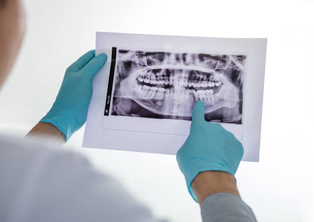
An X-ray does not show the nerve itself like a photograph would. It shows the space where the pulp sits, along with the surrounding tooth structure and bone.
In a healthy tooth, the pulp chamber and root canals appear as darker channels inside the tooth. Deep decay, a large filling near the pulp, or a shadow around the root tip may suggest irritation or infection. Those findings still need to be interpreted along with symptoms and testing.
During root canal treatment, the dentist removes diseased or dead pulp tissue from the chamber and canals, cleans the inside of the tooth, and seals it. Many patients first understand what a tooth nerve looks like only after seeing diagrams or clinical images during treatment or while researching a root canal.
Sometimes, yes, but usually not in the dramatic way people imagine. A deep cavity, broken tooth, or traumatic injury can expose the pulp. When that happens, it may look like a small red or bleeding point inside the tooth.
Still, many people who say the nerve is exposed are actually dealing with dentin exposure or gum recession. Those problems can cause sharp sensitivity without the pulp being directly visible. That difference matters because the right treatment may be very different.
If a tooth is fractured, bleeding from the center, or causing severe lingering pain, prompt dental care is the safer choice. Waiting can allow a manageable problem to become more complicated.
Some symptoms call for faster action. Emergency dentistry can provide same-day care for severe pain, swelling, or dental injuries. Facial swelling, fever, trouble swallowing, or difficulty opening the mouth should be assessed urgently.
Other reasons to arrange prompt care include severe throbbing pain, pain that wakes you from sleep, a broken tooth with visible inner tissue, or a tooth that suddenly changes color after trauma. Even without dramatic symptoms, recurring sensitivity or pain with biting should not be ignored.
General information can help you understand what may be happening, but it cannot confirm whether a tooth needs a filling, pulp cap, root canal treatment, crown, or extraction. That decision depends on an exam, imaging, and the condition of the tooth as a whole. If you need help with pain now, see our guide to toothache relief.
On the surface, this sounds like a simple anatomy question. In practice, it is often a pain question, a fear question, or a trust question. People want to know what is inside the tooth because they are trying to understand what has gone wrong and whether it can be fixed.
The honest answer is less theatrical than the popular image and more useful. A tooth nerve is not a lone wire. It is part of a small living organ inside the tooth, vulnerable to pressure, bacteria, cracks, and time.
That understanding can make dental decisions feel less mysterious. If a tooth has started sending stronger signals, or has gone strangely quiet after an injury, a dentist can help interpret those clues before the problem turns into an emergency. If fear is part of what is holding you back, learn how to reduce dental anxiety.
If you think you may need root canal treatment, Smileology can help. We have dental offices in Niceville, Miramar Beach, and Crestview, so you can schedule your evaluation at the location that's most convenient for you. Call our Niceville office at (850) 897-4488, our Miramar Beach office at (850) 424-7887, or our Crestview office at (850) 331-3392 to get started.
Not usually. When visible, it usually looks more like soft tissue inside the pulp space than a neat string or wire.
Not exactly. The nerve is part of the pulp, and the pulp also contains blood vessels and connective tissue.
Usually no. It is normally hidden beneath enamel and dentin. It may become visible if a tooth is badly decayed, fractured, or opened during treatment.
No. Tooth pain can come from several causes, including sensitivity, cracks, decay, gum recession, or pulp inflammation that may or may not be reversible. A dental evaluation is needed to tell the difference.
Healthy pulp tissue often appears pink to red because it contains blood vessels. Diseased or dying tissue may look darker or less uniform.